Beveled Osteotomies in Lateral Orbitotomy Using a Customized Rotating Bone Saw for Orbital Neoplasms

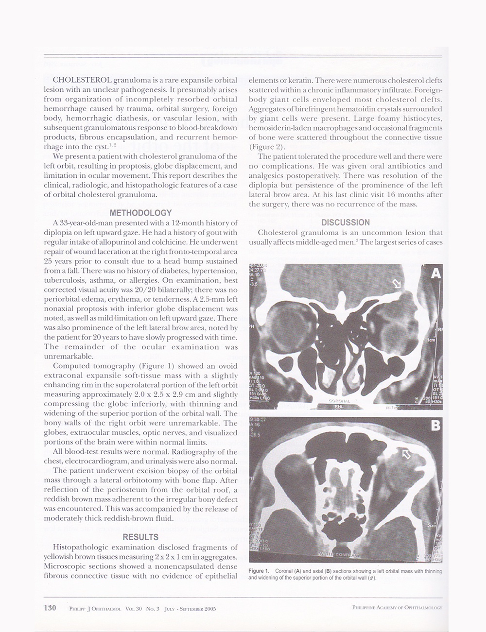
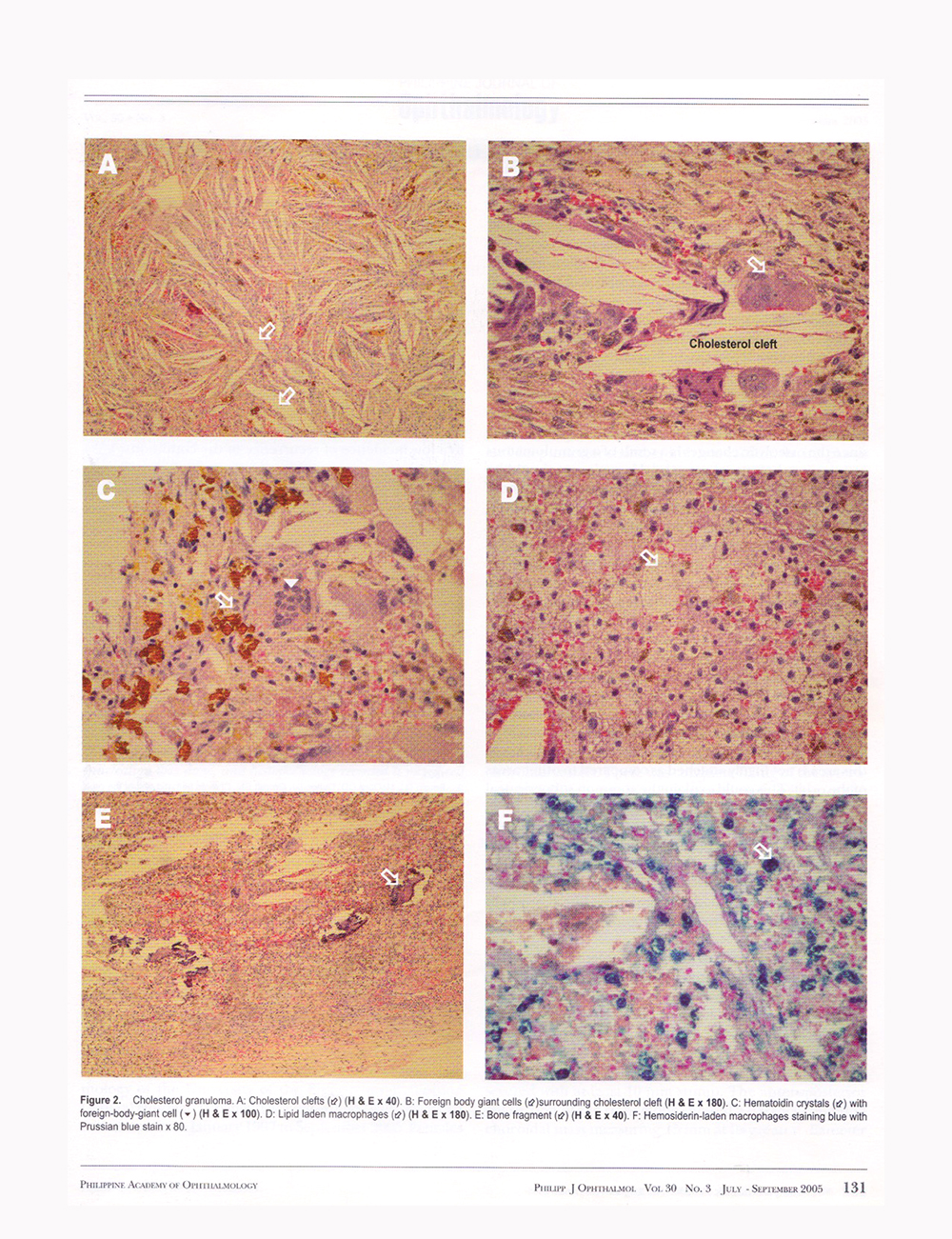
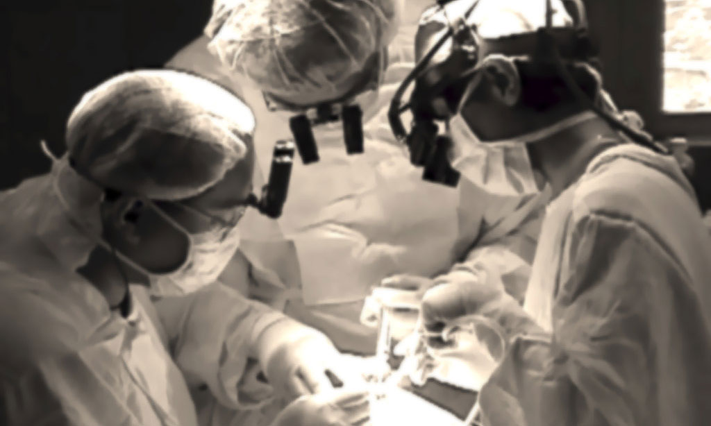
This study aims to develop a novel method of beveled osteotomy for lateral orbitotomy using a customized 21-mm stainless steel rotating saw in lateral orbitotomy and to evaluate the outcome of a novel beveled osteotomy in lateral orbitotomy. This article presents a case series (19 orbits from 18 patients) of lateral orbitotomies for excision biopsy of orbital neoplasms, over a 10-year period (from September 2001 to October 2011). It is a retrospective observational study. The surgeries were performed under the primary service of one surgeon (M. D. D. S.), the author of this study. All patients were treated via beveled osteotomies in lateral orbitotomy using stainless steel, 21 mm diameter, customized rotating bone saw. Preoperative and postoperative measurements were tabulated and statistically analyzed. The case series demonstrated that beveled osteotomies in lateral orbitotomy using stainless steel, 21 mm diameter, customized rotating bone saw was technically possible and provided access to lateral subperiorbital, peripheral, and central surgical spaces. The exposure was ample for excision biopsy of all neoplasms in this study. No patient needed the use of miniplate hardware in repositioning the lateral orbital wall nor complained of a palpable deformity of the lateral orbital wall. The wound healing was rapid, with minimal tissue distortion or scars. There were two patients who developed skin burns, but neither required cosmetic surgery to correct scarring from the burn. It was concluded that the modified technique of beveled osteotomies in lateral orbitotomy provides excellent access to the lateral subperiorbital, peripheral, and central surgical spaces. The exposure was adequate for excision biopsy of all neoplasms in this study. The technique promotes osseous union without the use of miniplate hardware. The use of a stainless steel 21 mm diameter customized rotating bone saw facilitated the successful outcome of the beveled technique.

Download PDF

Cholesterol Granuloma of the Orbit

Title: Cholesterol Granuloma of the Orbit

Published: Philippine Journal of Ophthalmology. Volume 30, No. 3 (July-September) 2005: pp 129-132

Objective:
To report a patient with cholesterol granuloma and describe the distinct clinical, radiologic, and histopathologic findings.

Methods: This is a case report.

Results:
A 33-year-old man presented with a 12-month history of diplopia on left upward gaze. He underwent repair of wound laceration at the right fronto-temporal area 25 years prior to consultation due to a head bump sustained in a vehicular accident. Examination revealed nonaxial proptosis, inferior globe displacement, and mild limitation on left upward gaze. Orbital imaging revealed an ovoid extraconal expansile soft-tissue mass in the left orbit, slightly compressing the globe inferiorly, and thinning and widening of the superior portion of the orbital wall. Excision biopsy of the orbital mass was done through a lateral orbitotomy with bone flap. Histopathology revealed characteristic features of cholesterol granuloma including abundant cholesterol clefts, foreign-body giant cells, lipid-laden histiocytes, and hemosiderin macrophages with absence of epithelial components.

Conclusion:
Orbitofrontal cholesterol granulomas have typical clinical, radiologic, and histopathologic features. Surgical incision has a high success rate with low incidence of recurrence.

The document pages (in jpg) containing the full article are below this post.

cholesterolgranuloma-p1 cholesterolgranuloma-p2 cholesterolgranuloma-p3 cholesterolgranuloma-p4

(end of document)

The Micro-Reflux Test – A New Test to Evaluate Nasolacrimal Duct Obstruction

Title: Micro-Reflux Test – A New Test to Evaluate Nasolacrimal Duct Obstruction

Published: Ophthalmology (USA). Volume 106, No. 12 (December) 1999 pp: 2319-2321

Objective: The authors describe a new and simple procedure, the Micro-Reflux Test (MRT), to screen for primary acquired nasolacrimal duct obstruction (PANDO). This study sought to determine the reliability of this new test in the diagnosis of complete nasolacrimal duct obstruction.

Design:
A nonrandomized, prospective, self-controlled comparative case series.

Participants: Two hundred eyes of 100 patients with documented unilateral complete PANDO were examined.

The document pages (in jpg) containing the full article are below this post.

Micro-Reflux-Test1 Micro-Reflux-Test2

Micro-Reflux-Test3

(end of document)

Endoscopic Laser-Assisted Dacryocystorhinostomy

Title: Surgical Technique of Endoscopic Laser-Assisted Dacryocystorhinostomy

Published. Operative Techniques in Oculoplastic, Orbital, and Reconstructive Surgery. Volume 1, No. 2 (December) 1998: pp 66-72

Caldwell described the endonasal approach to dacryocystorhinostomy (DCR) in 1893, but this approach was abandoned because of the technical complexities of the procedure. With the advent of endoscopy, there has been a renewed interest in the endonasal approach to DCR. Endoscopic laser-assisted DCR (ELA-DCR) has been the subject of much interest since its introduction in 1990 by Massaro et al. Despite its advantages, ELA-DCR has failed to become for widely accepted, clinical data have been sparse, and the effectiveness of this procedure has not been fully evaluated. The success rate of ELA-DCR in published small-series studies has ranged from 68.4% to 100%. More recently, Hartikainen et al compared the success rate and operative time between ELA-DCR and external DCR (EXT-DCR). In that report, the success rate 1 year after surgery was 91% for EXT-DCR and 63% for ELA-DCR. Their study also showed the operative time for ELA-DCR was three times shorter than that for EXT-DCR, with an average duration of 23 and 78 minutes respectively.

The document pages (in jpg) containing the full article are below this post.


dacryocystorhinostomy-1

dacryocystorhinostomy-2

dacryocystorhinostomy-3

dacryocystorhinostomy-4

dacryocystorhinostomy-p5

dacryocystorhinostomy-p6

dacryocystorhinostomy-p7

(end of file)


Diabetic Retinopathy in Non-Insulin Dependent Diabetes Mellitus

Title: Diabetic Retinopathy in Non-Insulin Dependent Diabetes Mellitus

Published. Philippine Journal of Ophthalmology. Volume 23, No. 3 (July-September) 1998: pp 116-120

Purpose: To identify the factors associated with the onset and progression of diabetic retinopathy.

Methods: One hundred fifteen (115) eyes of 59 patients were thoroughly examined for diabetic retinopathy. Baseline laboratory examinations were also performed and correlated with the ocular findings.

Results: Male sex, duration of the disease, fasting blood sugar, glysosylated hemoglobin and glucosuria were positively associated with diabetic retinopathy.

Conclusion: The importance of glycemic control on the progression of diabetic retinopathy is evident and therapeutic compliance guided by laboratory examinations is recommended.

The document pages (in jpg) containing the full article are below this post.

diabetic-retinopathy1 diabetic-retinopathy2 diabetic-retinopathy3 diabetic-retinopathy4 diabetic-retinopathy5(end of document)